Diagnosing pediatric liver disease requires a comprehensive, stepwise approach that combines clinical assessment, laboratory testing, imaging, and in many cases specialist investigations including genetic testing and liver biopsy.
Clinical history begins with a detailed account of the pregnancy, delivery, and neonatal period — including any antenatal infections, maternal medications, birth complications, or neonatal illness. A thorough family history explores whether any relatives have liver disease, metabolic conditions, or unexplained childhood deaths. The child's developmental history, feeding patterns, growth trajectory, and any medications or supplements they take are all relevant.
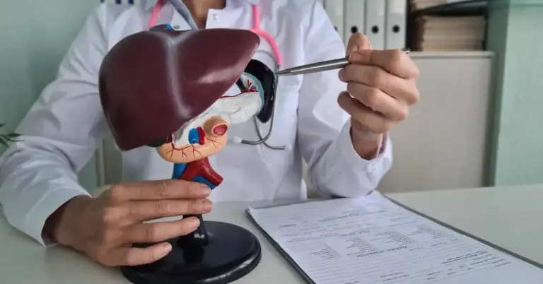
Physical examination assesses jaundice, the size and texture of the liver and spleen, the presence of ascites or abdominal distension, skin findings (spider angiomas, palmar erythema, excoriation from itching), growth parameters, and any extra-hepatic features suggesting specific diagnoses such as Alagille syndrome or Wilson's disease.
Blood tests provide the biochemical foundation of diagnosis. Liver function tests including ALT, AST, ALP, GGT, bilirubin (fractionated into direct and indirect components), albumin, and INR assess the degree of liver injury and synthetic function. A split bilirubin is particularly important in infants an elevated direct (conjugated) bilirubin always requires investigation regardless of the total bilirubin level.
Specific diagnostic blood tests are chosen based on the clinical picture. In suspected biliary atresia GGT level (characteristically very elevated), HIDA scan, and ultrasound. In suspected Wilson's disease serum ceruloplasmin, 24-hour urine copper, and slit-lamp examination. In alpha-1 antitrypsin deficiency alpha-1 antitrypsin level and phenotype. In autoimmune hepatitis ANA, ASMA, anti-LKM1, and immunoglobulin levels. In metabolic disorders plasma amino acids, urine organic acids, and specific enzyme assays.
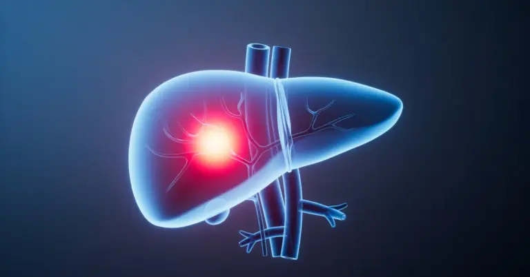
Liver ultrasound is the primary imaging tool in children it is safe, radiation-free, and provides excellent initial information about liver size, texture, bile duct anatomy, and portal blood flow. It is the first-line investigation for virtually all suspected paediatric liver conditions.
MRCP (MR cholangiopancreatography) provides detailed imaging of the bile duct system without radiation and is used to evaluate bile duct anatomy in conditions such as PSC, Alagille syndrome, and choledochal cysts.
FibroScan — measuring liver stiffness as a proxy for fibrosis is increasingly used in older children and adolescents. It is non-invasive, painless, and provides quantitative fibrosis staging.
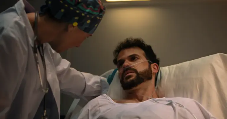
Liver biopsy remains important in paediatric hepatology providing definitive histological information about the degree of inflammation, fibrosis, and the specific pattern of injury that helps confirm diagnoses. In children with coagulopathy, the procedure is performed via the transjugular route. Under general anaesthesia in younger children.
Genetic testing has been transformed by next-generation sequencing technology. Gene panels covering dozens of paediatric liver disease genes can be tested simultaneously from a single blood sample, and in complex or undiagnosed cases, whole-exome or whole-genome sequencing can identify rare mutations that no targeted test would have found.
Intraoperative cholangiogram is performed during surgical exploration in suspected biliary atresia it is the definitive diagnostic step that confirms bile duct obliteration and triggers the Kasai procedure.